home
scroll down
view in full screen for best experience.

Introduction

Sound is simply moving air—compressed and rarefied molecules travelling in waves. When we produce or perceive a sound, we are really detecting patterns in air pressure changes over time.
These physical properties shape two key aspects of sound perception:
  • Pitch (frequency): Lower frequencies have more spread-out compressions and produce deeper pitches. Higher frequencies have closer compressions and create higher pitches.
  • Volume (amplitude): The amplitude of the wave determines loudness — low amplitude gives us quiet sounds, high amplitude gives us loud ones.
Humans can detect an impressive range of frequencies, from 20 Hz to 20,000 Hz (a modest range compared to other animals).

For example, bats and dolphins detect ultrasonic frequencies above 100,000 Hz! Elephants communicate using infrasonic rumbles below 20 Hz! Wow, the range of nature <3

The simple physics of sound is only the starting point. The real marvel is how the ear, with its delicate membranes, bones, and sensory cells, transforms these air vibrations into neural signals the brain then interprets as speech, music, or environmental cues.


In the chapters ahead, we’ll trace this transformation step by step.

The Auditory System

How is the ear built to capture and convert sound?
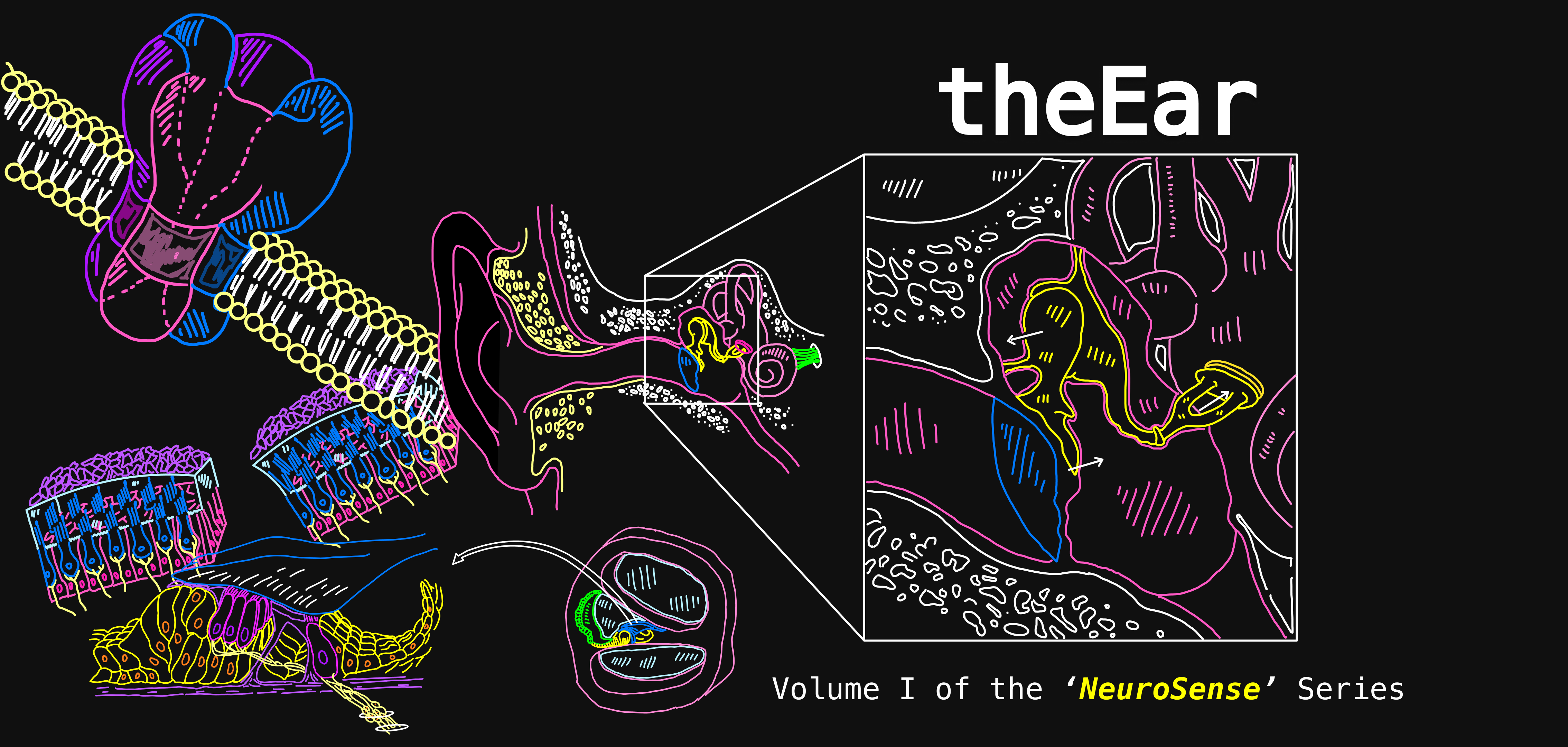
The auditory system works in 3 main stages.
  1. The Outer Ear
    Pinna: The visible part of the ear. Its curved, funnel-like shape helps capture sound waves and direct them inward.

    ‍• Auditory canal: A narrow tube that carries the waves toward the eardrum, concentrating and guiding them efficiently.


  2. The Middle Ear
    Eardrum (tympanic membrane): A thin, flexible membrane sitting at the end of the auditory canal. It vibrates when struck by sound waves.

    Ossicles: Three tiny bones that amplify the vibrations and pass them along. This is where air vibrations are converted into mechanical energy. 


  3. The Inner Ear
    Cochlea: A spiral, fluid-filled structure where mechanical vibrations become fluid waves. At this point, the wave ceases to be an air/mechanical wave and becomes a fluid one. 

    Auditory-vestibular nerve: Sends the information from the cochlea to the brain, where sound is recognised and interpreted.
let’s take a closer look!

Middle Ear

The ossicles form a chain of vibration from the eardrum to the inner ear.
  • Malleus (latin for hammer): The first place a wave is going to make contact with is our eardrum. This causes the malleus bone to start tapping/hammering on the next bone in the chain. 
  • Incus (latin for anvil): Receives the malleus’s force and transfers it onward.
  • Stapes (latin for stirrup): Horseshoe/stirrup shaped, the stapes presses against the oval window of the cochlea, passing the vibrations into the inner ear’s fluid.
Recap: Air enters → vibrates the eardrum → ossicles amplify and relay the motion → cochlea begins to vibrate.

Inner Ear

At the heart of the inner ear lies the cochlea. When the ossicles press against it, their mechanical force turns into a fluid wave.
If we were to take a cross-section of the cochlea, we would see it’s composed of 3 fluid-filled chambers:
  • Scala vestibuli (top chamber)
  • Organ of Corti (middle layer): A delicate, specialised structure that transduces vibrations into neural signals. 
  • Scala tympani (bottom chamber)
Together, these chambers allow fluid waves to move in carefully controlled patterns. 
Think of it like a balloon with one thinner patch in its surface. As the balloon fills with fluid and pressure builds inside, that weaker spot flexes outward, releasing strain and equalising the system.

Inside the Cochlea

To understand how the cochlea works, it helps to imagine it uncoiled as a straight tube. 

The cochlea is set up to detect sounds at different frequencies


Along its length runs the basilar membrane, which separates the scala vestibuli and scala timpani.

The basilar membrane isn’t uniform. Each section is tuned to a particular frequency, meaning it vibrates most strongly at that pitch. Mechanical properties determine which frequencies the basilar membrane is sensitive to.


So, the position along the cochlea determines how sensitive that part of the cochlea is to different sound frequencies.


In this way, the cochlea acts like a natural frequency analyser, translating the physical properties of sound into representative neural activity that the brain can interpret.

  • High-frequency sounds (i.e birds chirping) create strong vibrations near the base of the cochlea, but fade quickly.
  • Low-frequency (i.e. bass drum) travel further along the basilar membrane, peaking near the apex of the cochlea.
This spatial organization is called tonotopy (more on that later)—and it’s what allows us to distinguish between the vast range of pitches we hear every day.

Inside the Organ of Corti

How do vibrations in the cochlea turn into electrical signals?

The answers lies in the Organ of Corti.


The Organ of Corti (as a reminder) is a delicate, specialised structure that sits on top of the basilar membrane.

  • As fluid waves move through the cochlea, the basilar membrane vibrates up and down.

  • Resting above it is the tectorial membrane, a gelatinous structure that does not move much in response to these waves.
Note: Not to be confused with the tympanic membrane (our eardrum), which vibrates at the very start of the hearing pathway.

  • This difference in motion causes hair cells in between to be pushed against the tectorial membrane, deflecting their tiny projections (stereocilia).

    Stereocilia
    are stiff, toothpick-like structures on hair cells that bend in response to vibration.

Hair Cells

Within the Organ of Corti, we have two kinds of sensory receptors—outer and inner hair cells.
Together, they detect vibrations in the basilar membrane and begin the process of turning sound into neural signals.
  • Outer hair cells: Their stereocilia are embedded in the tectorial membrane, so they are directly deflected as the basilar membrane moves.
  • Inner hair cells: Their stereocilia float freely in the fluid. They respond to the overall fluid motion rather than direct contact.
Note: All hair cells are part of the Organ of Corti. While we often describe them separately as “inner” and “outer” hair cells, they together make up this specialised structure.
Hair cells themselves are not neurons because they don’t fire “all-or-nothing” action potentials.

Instead, they release neurotransmitters in a graded response, proportional to the frequency and amplitude of the incoming sound.

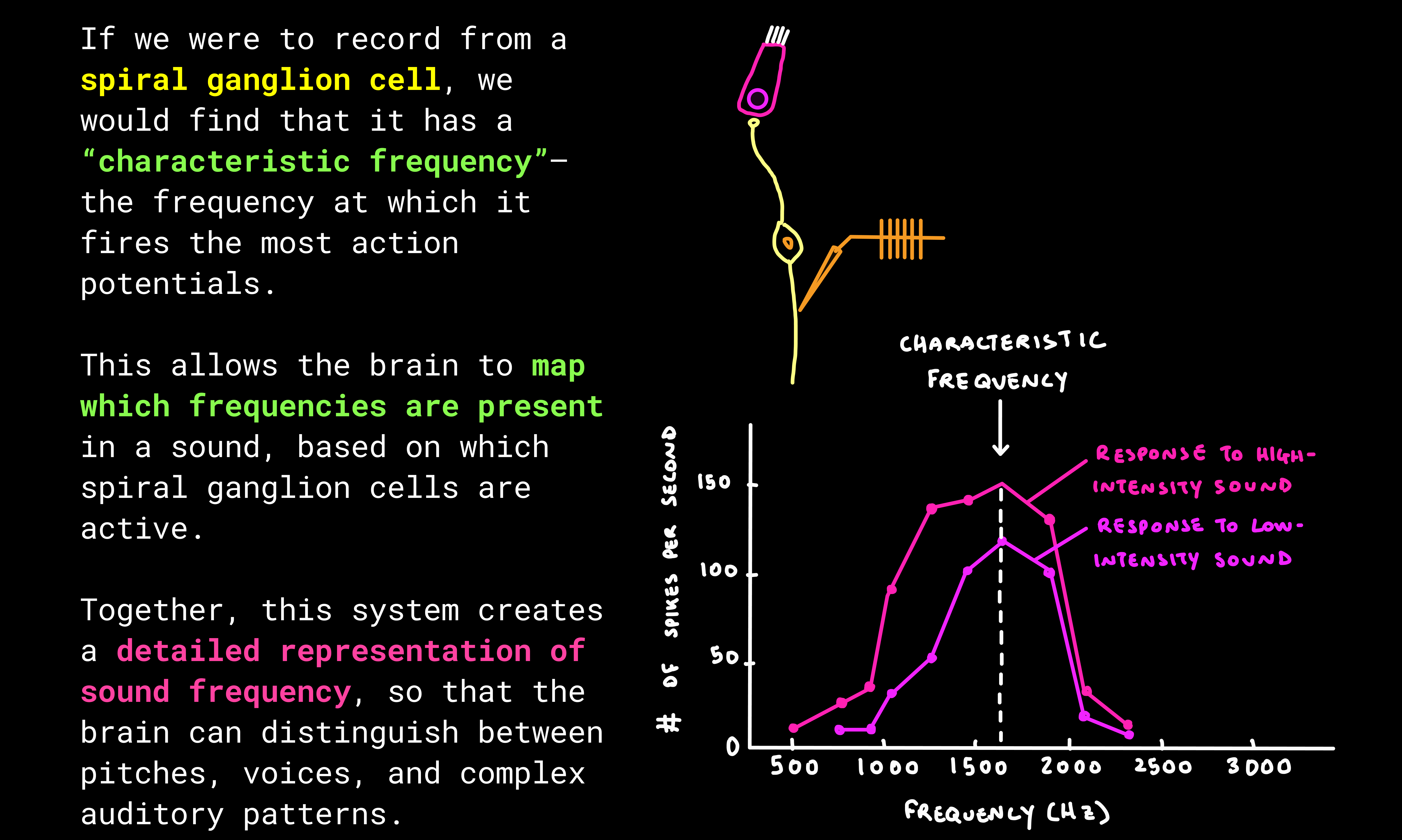
The first true neurons in this pathway are the spiral ganglion cells, which “listen” to the hair cells. Once activated, they fire action potentials and bundle their axons together to form the auditory nerve, carrying sound information to the brain.

Transduction

Below is a step-by-step process on how mechanical vibrations are converted into electrical signals that the brain can interpret.
Resting State
When hair cells are at rest, the basilar membrane is still and the stereocilia are upright. Only a few ion channels are open, so the cell sits near its resting potential.

Step 1: Deflection of Stereocilia
Each stereocilium is connected to its neighbour by tip-link proteins, so when one bends, the entire bundle shifts together.

  • As the basilar membrane vibrates, stereocilia bend toward one direction, opening mechanically-gated potassium (K+) channels.

  • When they bend the other way, the channels close.
Step 3: Neurotransmitter Release
Depolarisation opens voltage-gated calcium (Ca2+) channels.

  • Ca2+ rushes in, triggering vesicles to fuse with the membrane.
  • These vesicles release neurotransmitter onto the spiral ganglion cells.

    The release is graded—always present at some level, but stronger or weaker depending on the frequency and intensity of the sound.
  • Spiral ganglion cells listen to the graded messages of the hair cells and decide whether or not they will fire a binary action potential. They pass this chemical signal into the auditory nerve and onward to the brain.

Recap: Sound wave enters ear basilar membrane vibrates stereocilia bend K⁺ channels open/close depolarisation / hyperpolarisation of hair cells Ca²⁺ influx neurotransmitter release spiral ganglion fires auditory nerve brain.
Differences Between Outer and Inner Hair Cells
Although both types of hair cells detect motion in the cochlea, they connect to spiral ganglion cells in very different ways.
  • Many outer hair cells converge onto a single spiral ganglion cell. This means their information is pooled together before being passed on.
  • Conversely, we have the opposite arrangement for inner hair cells. One inner hair cell communicates with many spiral ganglion cells. This spreads its information more widely through the pathway.
Why the Difference?
Outer hair cells actually outnumber inner hair cells 3:1.

(Thus, there’s more outer hair cells than inner hair cells)

Yet, most spiral ganglion cells are connected to inner hair cells. Only a small fraction listen to the outer ones.

This is because the two groups encode different aspects of sound.
  • Inner hair cells: Specialised for frequency (pitch) detection.
  • Outer hair cells: More sensitive to amplitude (volume) intensity.
While amplitude helps us gauge how strong a sound is, frequency is more critical for distinguishing and recognising sounds.

e.g., It’s the change in pitch that lets us tell voices apart or identify a melody.

Hearing Loss

Hearing loss can result from many different causes, depending on where the auditory pathway is disrupted.

Broadly, hearing loss is divided into conductive deafness and sensorineural deafness.

Conductive Deafness
This occurs when sound waves are blocked from reaching the cochlea.
  • Common causes: Ear infections, fluid buildup, damaged/calcification of the ossicles, or ruptured ear drum.

    In these instances, mechanical vibrations cannot be transmitted effectively through the middle ear.
  • Hearing aids are often effective here since they amplify incoming sound waves. Surgery may also help repair ossicles or eardrum damage.

Sensorineural Deafness
This occurs when the cochlea or auditory nerve is damaged.

One of the most common causes is malfunctioning hair cells in the cochlea. In these cases, cochlear implants can help.
  • A cochlear implant works by bypassing the damaged hair cells entirely.

    It acts like an artificial basilar membrane, directly stimulating the auditory nerve with electrical signals instead of relying on mechanical-to-neural conversion.

These can recover up to ~90% of hearing ability!
However, cochlear implants are not helpful for every type of hearing loss.
  • Auditory nerve damage: If the nerve itself is damaged, bypassing the hair cells is not enough, since the pathway to the brain is interrupted.
Mixed Hearing Loss
Sometimes both conductive and sensorineural components are present. For example, someone might have ossicle calcification and cochlear hair cell damage. Procedures may then involve a combination of surgery, hearing aids, or implants, depending on the primary cause.

Cochlear Amplification

Outer hair cells also amplify sound.

On their surface, they contain a special protein called prestin, a voltage-sensitive motor protein.

How It Works
  • When the hair cell depolarises, prestin contracts, pushing the cell against the tectorial membrane and boosting the effect of the vibration.

  • When the hair cell hyperpolarises, prestin relaxes, allowing the cell to expand and pull away, further enhancing the contrast.
This rapid expansion and contraction acts like a biological amplifier, strengthening the response to sound waves already being detected by the stereocilia.

Why Does This Matter?
Cochlear amplification allows us to detect sounds at very low intensities that would otherwise be too faint to perceive.

It fine-tunes our sensitivity to quiet sounds and subtle differences in pitch.

Clinical note: The drug furosemide can inhibit prestin proteins, removing this amplification mechanism. As a result, the ear becomes less sensitive, particularly to low-intensity sounds.

Audition Pathway

The journey of sound from ear to brain begins with our lovely spiral ganglion cells, the first true neurons of the auditory nerve.
Reminder: These cells “listen” to the hair cells in the cochlea and then pass the signal onward.

Step I: Cochlear Nucleus

  • Each auditory nerve projects to the ventral cochlear nucleus on the same side of the brainstem.
  • At this stage, information is still unilateral. e.g., input from the left cochlea comes from the left auditory nerve and hits the left ventral cochlear nucleus (and vice versa).

Step II: Superior Olives

  • From the cochlear nucleus, signals split and become bilateral.
  • The cell in the central cochlear nucleus sends information to both the right and the left superior olives.
  • The superior olives are a cluster of brainstem neurons that compare input from both ears to determine sound direction.
This comparison is essential for sound localisation
The ventral cochlear nucleus still only gets input from one side.

In contrast, the superior olive receives input from both ears, allowing it to compare signals.

This mechanism, interaural time difference (ITD), is key for localising sound left vs. right.

Other processes, like interaural intensity differences, help with front vs. back localisation.

Step 3: Inferior Colliculus

Step 4: Thalamus (MGN)

Step 5: Auditory Cortex (A1)

Finally, the signal arrives at the primary auditory cortex (A1), located just below the lateral fissure. Here, sound is consciously processed, recognised, and interpreted.

Recap: Cochlea (hair cells → spiral ganglion) → Ventral Cochlear Nucleus → Superior Olive (binaural comparison) → Inferior Colliculus → Thalamus (MGN) → Auditory Cortex (A1)

Tonotopy

The frequency map of the cochlea is not lost as signals travel upward, it is preserved all the way to the brain.

Remember, the basilar membrane is arranged tonotopically. Different regions respond best to different frequencies.

Hair cells sitting at each point along the membrane inherit this tuning.

!Reminder!
Hair cells maintain frequency-specific pathways as the signal ascends through the brainstem, thalamus, and finally into A1.

This preserves the frequency-specific coding in A1.
In A1, the map is laid out spatially:
  • Posterior regions respond most strongly to high frequencies.
  • Anterior regions respond most strongly to low frequencies.
This means the auditory cortex itself contains a physical “map” of pitch, allowing the brain to separate and organise complex sounds like speech and music.
Beyond the Primary Auditory Cortex...
A1 sorts sounds by frequency, timing, and pattern. But meaning comes from what happens next: connections to association areas like the prefrontal cortex and the limbic system. These connections allow us to recognise voices, understand language, and process music with memory and emotion.

BUT, that’s a story for another time...another series?

The Vestibular System

The vestibular system is what allows us to maintain balance, know where we are in space, and sense how our body is moving relative to gravity.


Next to the cochlea lies the vestibular labyrinth, made up of three semicircular canals.

Unlike the cochlea, these structures don’t respond to sound. Instead, they detect movements of the head and make compensatory movements accordingly.

Each canal is oriented in a different plane:
Up/Down. Left/Right. Tilt/Rotation.

This is why there are three: together, they cover all possible directions of head movement.

Anatomy of the Vestibular System

The core structure is the otolith organ.

The otolith organ includes the utricle and saccule. Inside are hair cells whose stereocilia extend into a gelatinous layer.

As the head moves, the jelly-like layer sloshes, bending the stereocilia back and forth.
The otolith organs are especially tuned to detect acceleration.
Why Is This Important?
The vestibular system drives the Vestibulo-Ocular Reflex (VOR), which automatically adjusts our eye position when our head moves.

This keeps visual input steady on the retina so we can stay focused on objects even while moving.

When the vestibular system malfunctions, prediction and compensation break down, leading to motion sickness.

Summary

Wow, that was a lot of information. Let's recap!

Closing Remarks


The auditory and vestibular systems remind us how extraordinary the brain is.
To hear the softess whisper, the pulse of music, or the rush of laughter; To stay upright or balanced on a moving bus—

These systems keep us grounded in the world and attuned to one another.

By peering more deeply into the ear, we gain another lens on perception itself: how the brain interprets vibration and motion in our daily lives.

My hope is that you see the elegance of neuroscience, and feel inspired to listen more closely (now that you know how to do it!)—to sound, to sensation, and to each other.

Stay tuned! The next neuroSense volume will turn from the ear to the eye, exploring how we perceive the world through sight.

thanks for reading!

amethyst

Key Terms

Not a neuroscientist? Don’t worry!
Below are definitions of common terms used throughout the volume.

Neurotransmitters
These are the brain’s chemical messengers. They carry signals between neurons across the synapse (the tiny gap between cells).

When an action potential reaches the end of an axon, it triggers the release of neurotransmitters. These molecules cross the synapse and bind to receptors on the next neuron, influencing whether that cell will fire its own signal.

Different neurotransmitters have different effects: some excite (make a neuron more likely to fire), others inhibit (make it less likely).
Depolarisation / Hyperpolarisation
Neurons maintain a difference in charge between the inside and outside of the cell. The inside is usually more negative than the outside (this is called the resting membrane potential).

Charged particles (ions) move according to gradients: positives are attracted to negatives, and vice versa. Uncharged particles follow different rules of diffusion, but this isn’t relevant here.

  • Depolarisation: The inside becomes less negative (more positive) moving closer to the threshold needed to fire an action potential. This increases the likelihood that an action potential will occur.

  • Hyperpolarisation: The inside becomes even more negative, moving further from the threshold. This reduces the chance that an action  potential will occur.
Mechanically Gated Channels
These are proteins in the cell membrane that act like doors, opening or closing in response to physical movement. Unlike other forms of channels (chemical or voltage-gated), they don’t rely on signals or electricity — they move when the membrane itself is pushed, stretched, or bent.

In the ear, bending the tiny hair-like stereocilia physically pulls these channels open, letting ions rush in and starting the process of sound transduction.

Voltage-Gated Channels
These proteins open or close in response to changes in electrical charge across the cell membrane. They are triggered when the neuron becomes more or less electrically polarised.

When the voltage shifts past a certain threshold, these channels snap open, allowing ions—like sodium (Na⁺) or calcium (Ca²⁺)—to rush in. This sudden flow of charge helps generate or propagate an action potential.

Voltage-gated channels are essential for rapid signalling in the nervous system: they detect a change in electricity, respond instantly, and help carry messages along neurons at remarkable speeds. In the ear, these channels help convert the graded signals from hair cells into full electrical impulses that travel along the auditory nerve to the brain.

Why Do We Need Channels?
Because the cell membrane (made of a phospholipid bilayer) is naturally impermeable to charged particles like ions. Water and small uncharged molecules can slip through, but ions can’t cross on their own. Channels create the necessary “gates” for them to pass through.