scroll down
view in full screen for best experience.

Introduction

Sound is simply moving air—compressed and rarefied molecules travelling in waves. When we produce or perceive a sound, we are really detecting patterns in air pressure changes over time.
Diagram showing sound wave propagation from a stereo speaker. Green dots represent air molecules alternating between compressed regions (closer together) and rarefied regions (spread apart). Below, a pink sine wave graphs air pressure over distance, with peaks aligned with compressed zones and troughs aligned with rarefied zones. One complete cycle is marked between consecutive peaks.

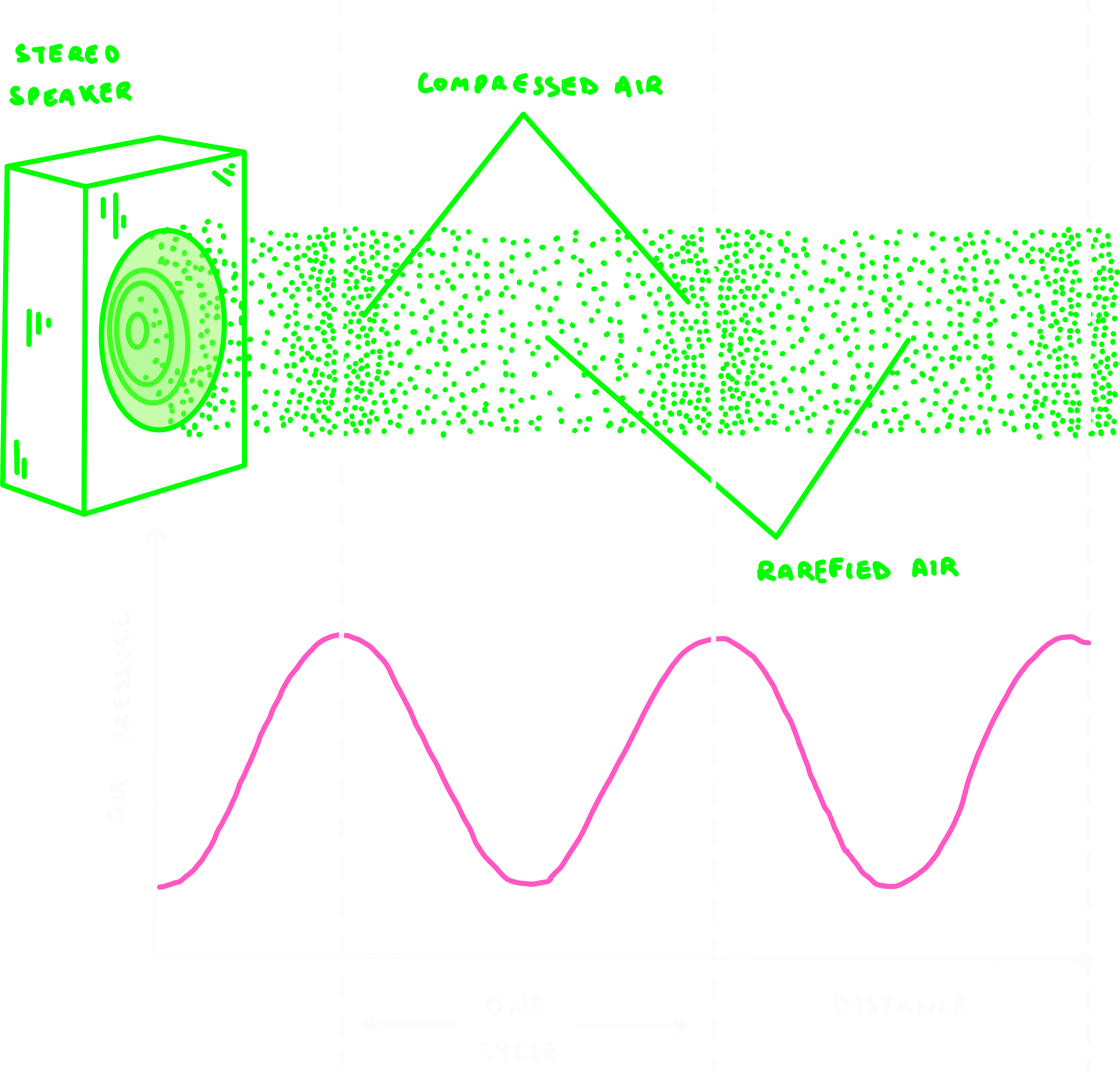
Compressed air: Regions of the wave where air molecules are closer together.

Rarefied air: Parts of the wave where air molecules are spread further apart.

There’s more pressure in compressed zones, and less pressure in rarefied zones. This alternation produces the wave-like structure of a sound wave.

A cycle is the distance between peaks or troughs of the wave.

These physical properties shape two key aspects of sound perception.
  • Pitch (frequency): Lower frequencies have more spread-out compressions and produce deeper pitches. Higher frequencies have closer compressions and create higher pitches.
Two side-by-side sine wave graphs comparing low frequency sound (fewer, wider waves) and high frequency sound (many compressed waves closely spaced together)
  • Volume (amplitude): The amplitude of the wave determines loudness — low amplitude gives us quiet sounds, high amplitude gives us loud ones.
Alt text option 1 (concise): "Two side-by-side sine wave graphs comparing low intensity sound (small amplitude waves) and high intensity sound (large amplitude waves)
Humans can detect an impressive range of frequencies, from 20 Hz to 20,000 Hz (a modest range compared to other animals).

For example, bats and dolphins detect ultrasonic frequencies above 100,000 Hz! Elephants communicate using infrasonic rumbles below 20 Hz! Wow, the range of nature <3

The simple physics of sound is only the starting point. The real marvel is how the ear, with its delicate membranes, bones, and sensory cells, transforms these air vibrations into neural signals the brain then interprets as speech, music, or environmental cues.
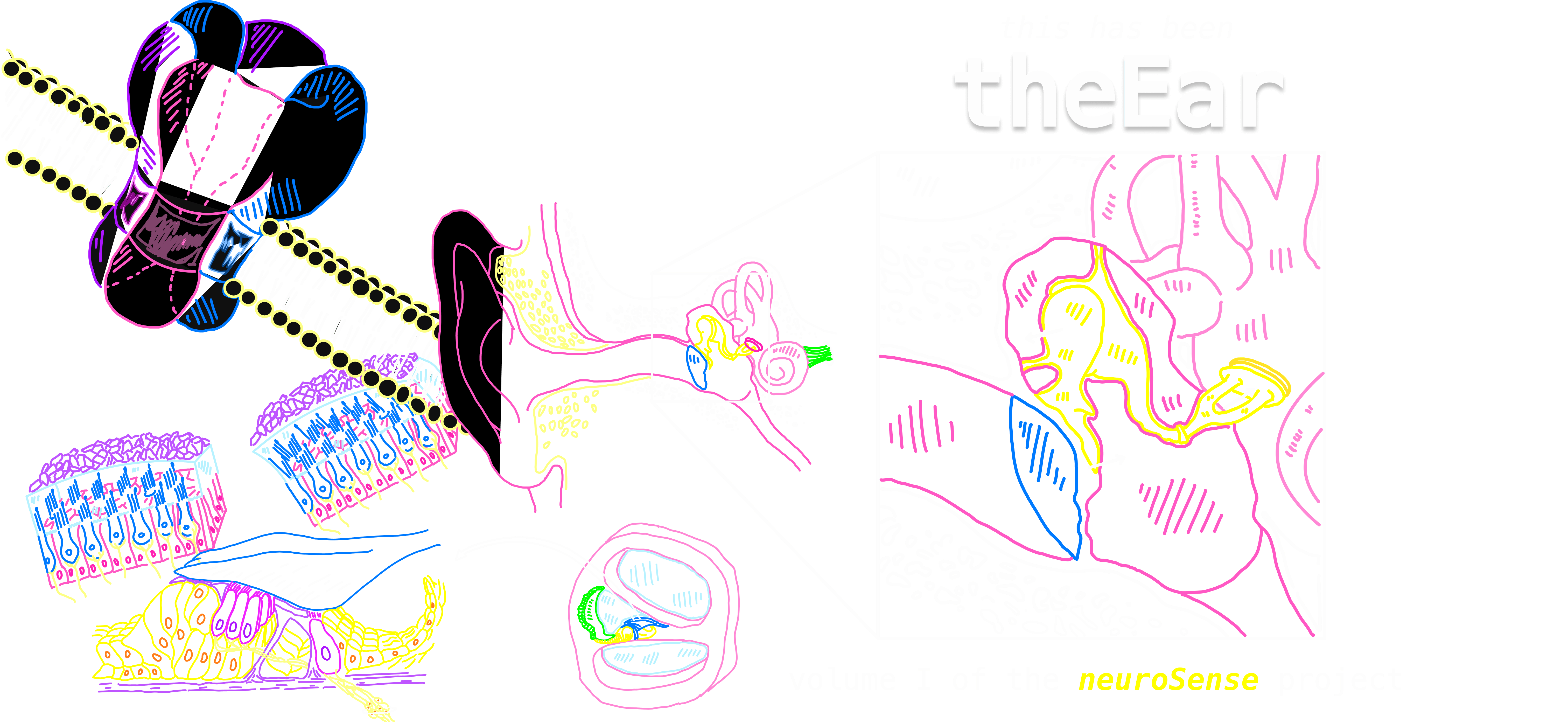
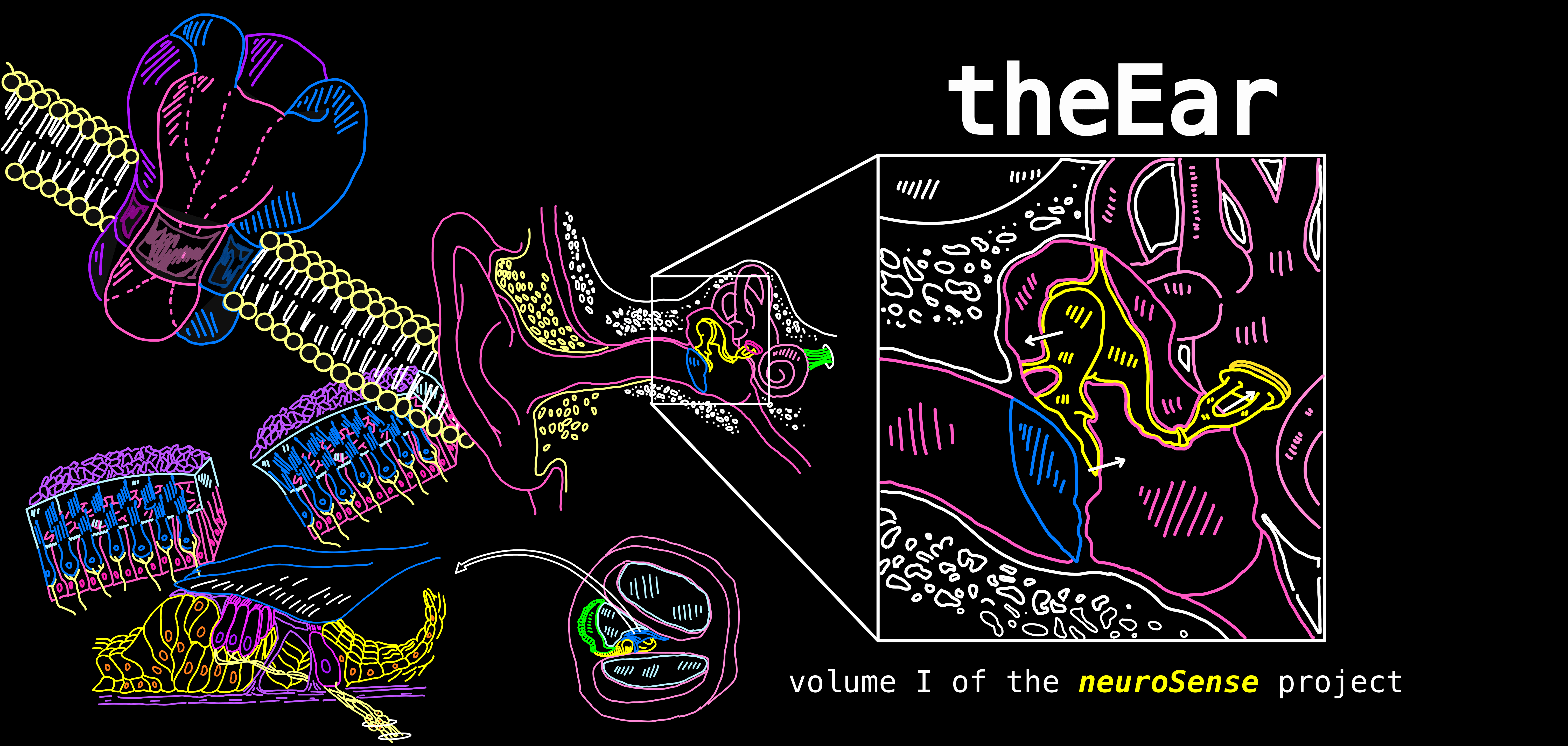

In the chapters ahead, we’ll trace this transformation step by step.

The Auditory System

How is the ear built to capture and convert sound?
Cross-sectional diagram of the human ear illustrating how sound is captured and converted. Labels identify key structures across the outer ear (pinna, auditory canal), middle ear (ossicles), and inner ear (oval window, cochlea, tympanic membrane/eardrum, auditory-vestibular nerve). Arrows indicate the path of sound through the three main stages of the auditory system.
The auditory system works in 3 main stages.
  1. The Outer Ear
    Pinna: The visible part of the ear. Its curved, funnel-like shape helps capture sound waves and direct them inward.

    Auditory canal: A narrow tube that carries the waves toward the eardrum, concentrating and guiding them efficiently.


  2. The Middle Ear
    Eardrum (tympanic membrane): A thin, flexible membrane sitting at the end of the auditory canal. It vibrates when struck by sound waves.

    Ossicles: Three tiny bones that amplify the vibrations and pass them along. This is where air vibrations are converted into mechanical energy. 


  3. The Inner Ear
    Cochlea: A spiral, fluid-filled structure where mechanical vibrations become fluid waves. At this point, the wave ceases to be an air/mechanical wave and becomes a fluid one. 

    Auditory-vestibular nerve: Sends the information from the cochlea to the brain, where sound is recognised and interpreted.
let’s take a closer look!

Anatomical diagram showing a cross-section of the ear with a magnified inset detail. The left side shows the overall ear structure in pink outline with yellow-highlighted ossicles. The right side presents a detailed close-up view of the middle ear cavity, clearly labeling the three tiny bones (malleus, incus, and stapes in yellow), the footplate of the stapes at the oval window, the cochlea (pink spiral structure), the auditory canal, the tympanic membrane in blue, and the Eustachian tube, illustrating how these structures connect to transmit sound vibrations.

Middle Ear

The ossicles form a chain of vibration from the eardrum to the inner ear.
  • Malleus (latin for hammer): The first place a wave is going to make contact with is our eardrum. This causes the malleus bone to start tapping/hammering on the next bone in the chain. 
  • Incus (latin for anvil): Receives the malleus’s force and transfers it onward.
  • Stapes (latin for stirrup): Horseshoe/stirrup shaped, the stapes presses against the oval window of the cochlea, passing the vibrations into the inner ear’s fluid.
Air enters vibrates the eardrum ossicles amplify and relay the motion cochlea begins to vibrate.

Pressure Regulation
The ear connects to the mouth and sinus cavity through the eustachian tube.

When air pressure outside changes, pressure can build in the middle ear.

When we swallow or yawn, our ears will pop!
This is our eardrum shifting to release the pressure.

Protective Muscles
There are 2 muscles within the middle ear:

  • Stapedius muscle
  • Tensor tympani muscle
They act as emergency stabilisers for the auditory system.
Cross-sectional anatomical diagram of the human ear showing the outer, middle, and inner ear structures. Key labelled structures include the ossicles (three small bones in yellow), labyrinth (in pink), oval window (yellow), round window (light pink), cochlea (green), tensor tympani muscle (pink), stapedius muscle (pink), and tympanic membrane/eardrum (blue). The eustachian tube connects the middle ear to the throat cavity.
Their role is to temporarily stiffen the ossicles when sound is dangerously loud.

e.g., At a loud concert, these muscles tense up to reduce the energy passed along, protecting the delicate cochlea.

These muscles are also activated when we speak, slightly dampening the sound of our own voice.

Inner Ear

At the heart of the inner ear lies the cochlea. When the ossicles press against it, their mechanical force turns into a fluid wave.
If we were to take a cross-section of the cochlea, we would see it’s composed of 3 fluid-filled chambers:
  • Scala vestibuli (top chamber)
  • Organ of Corti (middle layer): A delicate, specialised structure that transduces vibrations into neural signals. 
  • Scala tympani (bottom chamber)
Together, these chambers allow fluid waves to move in carefully controlled patterns. 
Anatomical diagram depicting the auditory system with a full ear cross-section and a magnified cochlear detail. The left portion of the image shows the middle and inner ear outlined in bright neon colors: the tympanic membrane in blue, the ossicles in yellow, the tensor tympani and stapedius muscles in pink, and the labyrinth and cochlea in pink outline. An arrow leads to the right side, which presents a detailed close-up cross-section of the cochlea, showing the scala vestibuli, scala media, and scala tympani in pastel tones, with labeled structures including Reissner’s membrane, the tectorial membrane in blue, the stria vascularis in green, the basilar membrane in pink, and the organ of Corti in yellow. The illustration emphasises how these connected structures participate in sound transduction.

Pressure Valves

For the cochlea to function, sound waves need a way in and a way out. Two small membranes act as pressure release valves.

  • Oval Window: The stapes presses against this membrane, transmitting vibrations into the scala vestibuli and setting the cochlear fluid in motion.
  • Round Window: A thin, flexible membrane at the other end of the cochlea. As fluid waves travel through the cochlea, the round window bulges outward to release the extra pressure.

    Without the round window, the incompressible fluid of the cochlea would have nowhere to move, and the stapes wouldn’t be able to set up waves effectively.
Think of it like a balloon with one thinner patch in its surface. As the balloon fills with fluid and pressure builds inside, that weaker spot flexes outward, releasing strain and equalising the system.

Inside the Cochlea

To understand how the cochlea works, it helps to imagine it uncoiled as a straight tube. 

The cochlea is set up to detect sounds at different frequencies
An illustrated diagram showing how the cochlea uncoils and how its internal structures are arranged. The top row depicts the cochlea in pink outline transitioning from its natural spiral shape to a fully uncoiled form. The bottom illustration shows the uncoiled cochlea in detail, labelling the oval window where the stapes footplate (shown in yellow) connects, the round window, and the three main fluid-filled chambers: scala vestibuli, scala media, and scala tympani (pink) The basilar membrane spans the length of the cochlea, widening toward the apex, with the helicotrema marked at the far end where the chambers connect. The diagram visualises how sound waves travel along the basilar membrane from base to apex

Along its length runs the basilar membrane, which separates the scala vestibuli and scala timpani.

The basilar membrane isn’t uniform. Each section is tuned to a particular frequency, meaning it vibrates most strongly at that pitch. Mechanical properties determine which frequencies the basilar membrane is sensitive to.


So, the position along the cochlea determines how sensitive that part of the cochlea is to different sound frequencies.

Diagram illustrating how the cochlea processes different sound frequencies through tonotopic organisation. The top panel shows the cochlea uncoiled into a tube, labeled with the stapes at the entrance, round window, perilymph fluid, basilar membrane running through the center, and helicotrema at the far end. The bottom three panels demonstrate the basilar membrane's changing properties: (a) shows the base as narrow and stiff for high frequency detection, (b) shows the apex as wide and floppy for low frequency detection, and (c) displays the complete frequency gradient with specific measurements from 500 Hz at the apex through 1 kHz, 2 kHz, 4 kHz, 8 kHz, to 16 kHz at the base, illustrating how maximum amplitude response varies by location along the membrane.

In this way, the cochlea acts like a natural frequency analyser, translating the physical properties of sound into representative neural activity that the brain can interpret.

  • High-frequency sounds (i.e birds chirping) create strong vibrations near the base of the cochlea, but fade quickly.
  • Low-frequency (i.e. bass drum) travel further along the basilar membrane, peaking near the apex of the cochlea.
This spatial organisation is called tonotopy (more on that later)—and it’s what allows us to distinguish between the vast range of pitches we hear every day.

Inside the Organ of Corti

How do vibrations in the cochlea turn into electrical signals?

The answers lies in the Organ of Corti.

Comprehensive anatomical illustration of the organ of Corti, the sensory organ within the cochlea responsible for detecting sound vibrations. The left panel shows a detailed side view with labeled structures: the tectorial membrane (blue) positioned above rows of hair cells, outer hair cells (three rows in yellow) and inner hair cells (one row in pink) arranged on the basilar membrane (purple baseline), stereocilia (hair-like projections) extending from the hair cells toward the tectorial membrane, the rods of Corti providing structural support, and nerve fibers (yellow) connecting the hair cells to the spiral ganglion and auditory nerve. The right panel shows a cross-sectional view of the entire cochlear duct with the organ of Corti (green) positioned within, and the central modiolus (yellow core) where nerve fibres converge.


The Organ of Corti (as a reminder) is a delicate, specialised structure that sits on top of the basilar membrane.

  • As fluid waves move through the cochlea, the basilar membrane vibrates up and down.

  • Resting above it is the tectorial membrane, a gelatinous structure that does not move much in response to these waves.
Note: Not to be confused with the tympanic membrane (our eardrum), which vibrates at the very start of the hearing pathway.

This difference in motion causes hair cells in between to be pushed against the tectorial membrane, deflecting their tiny projections (stereocilia).

Stereocilia
are stiff, toothpick-like structures on hair cells that bend in response to vibration.

Hair Cells

Within the Organ of Corti, we have two kinds of sensory receptors—outer and inner hair cells.
Together, they detect vibrations in the basilar membrane and begin the process of turning sound into neural signals.
  • Outer hair cells: Their stereocilia are embedded in the tectorial membrane, so they are directly deflected as the basilar membrane moves.
  • Inner hair cells: Their stereocilia float freely in the fluid. They respond to the overall fluid motion rather than direct contact.
Side-by-side comparison  illustrating the mechanical motion of the Organ of Corti during sound detection. The left panel depicts the structure at rest, with three outer hair cells (pink) and one inner hair cell (pink) positioned on the basilar membrane (cyan baseline), their stereocilia extending upward toward the tectorial membrane (blue), with the rods of Corti (purple) providing support and the modiolus (yellow) shown as the central axis. The right panel shows the same structure during sound-induced vibration, where the basilar membrane deflects upward (indicated by dashed line showing original position), causing the stereocilia to bend against the tectorial membrane, which triggers the transduction process that converts mechanical motion into neural signals.
Note: All hair cells are part of the Organ of Corti. While we often describe them separately as “inner” and “outer” hair cells, they together make up this specialised structure.
Hair cells themselves are not neurons because they don’t fire “all-or-nothing” action potentials.

Instead, they release neurotransmitters in a graded response, proportional to the frequency and amplitude of the incoming sound.

The first true neurons in this pathway are the spiral ganglion cells, which “listen” to the hair cells. Once activated, they fire action potentials and bundle their axons together to form the auditory nerve, carrying sound information to the brain.

Transduction

Below is a step-by-step process on how mechanical vibrations are converted into electrical signals that the brain can interpret.
Resting State
When hair cells are at rest, the basilar membrane is still and the stereocilia are upright. Only a few ion channels are open, so the cell sits near its resting potential.

Step 1: Deflection of Stereocilia
Each stereocilium is connected to its neighbour by tip-link proteins, so when one bends, the entire bundle shifts together.
  • As the basilar membrane vibrates, stereocilia bend toward one direction, opening mechanically-gated potassium (K+) channels.

  • When they bend the other way, the channels close.

Step 2: Ion Flow
Unlike in most of the central nervous system (CNS), the fluid around the hair cells — the endolymph — contains a high concentration of potassium.

Because the inside of the hair cell is relatively negative, positively charged ions are strongly driven to enter when channels open.

  • The influx of K+ ions depolarises the cell.
  • As the wave reverses, the channels close, and the cell becomes hyperpolarised again.
Diagram illustrating ion flow in cochlear hair cells. Three sequential panels on the left show mechanically gated channels (orange) on stereocilia opening and closing as the hair bundle deflects. The right panel shows a complete inner hair cell with stereocilia containing potassium ions (K+) in the endolymph above. When channels open, K+ ions enter, causing depolarisation. This triggers voltage-gated calcium channels (blue) to open at the cell base, allowing Ca2+ influx. Vesicles filled with glutamate neurotransmitter (green) are shown ready for release at the synapse with the spiral ganglion neurite below. The reticular lamina (purple) separates the endolymph (cyan) from the perilymph  surrounding the cell body (cyan). 

This rapid alternation encodes the timing and rhythm of the incoming sound wave.
Step 3: Neurotransmitter Release
Depolarisation opens voltage-gated calcium (Ca2+) channels.

  • Ca2+ rushes in, triggering vesicles to fuse with the membrane.
  • These vesicles release neurotransmitter onto the spiral ganglion cells.

    The release is graded—always present at some level, but stronger or weaker depending on the frequency and intensity of the sound.
  • Spiral ganglion cells listen to the graded messages of the hair cells and decide whether or not they will fire a binary action potential. They pass this chemical signal into the auditory nerve and onward to the brain.

Recap: Sound wave enters ear basilar membrane vibrates stereocilia bend K⁺ channels open/close depolarisation / hyperpolarisation of hair cells Ca²⁺ influx neurotransmitter release spiral ganglion fires auditory nerve brain.
Differences Between Outer and Inner Hair Cells
Although both types of hair cells detect motion in the cochlea, they connect to spiral ganglion cells in very different ways.
Illustration of auditory neural connectivity showing how hair cells transmit information to the brain. On the left, three outer hair cells (pink cell bodies with stereocilia) send efferent connections (yellow nerve fibres) that converge toward a single spiral ganglion. On the right, one inner hair cell (pink) with stereocilia extending upward sends multiple afferent nerve fibres (yellow) downward to synapse with a cluster of spiral ganglion cells.  The axons from these spiral ganglion neurons bundle together to form the auditory nerve (green fibers at bottom), which carries the encoded sound information to the brainstem and ultimately to auditory cortex.
  • Many outer hair cells converge onto a single spiral ganglion cell. This means their information is pooled together before being passed on.
  • Conversely, we have the opposite arrangement for inner hair cells. One inner hair cell communicates with many spiral ganglion cells. This spreads its information more widely through the pathway.
Why the Difference?
Outer hair cells actually outnumber inner hair cells 3:1.

Thus, there’s more outer hair cells than inner hair cells.

Yet, most spiral ganglion cells are connected to inner hair cells. Only a small fraction listen to the outer ones.

This is because the two groups encode different aspects of sound.
  • Inner hair cells: Specialised for frequency (pitch) detection.
  • Outer hair cells: More sensitive to amplitude (volume) intensity.
While amplitude helps us gauge how strong a sound is, frequency is more critical for distinguishing and recognising sounds.

e.g., It’s the change in pitch that lets us tell voices apart or identify a melody.

Hearing Loss

Hearing loss can result from many different causes, depending on where the auditory pathway is disrupted.

Broadly, hearing loss is divided into conductive deafness and sensorineural deafness.

Conductive Deafness
This occurs when sound waves are blocked from reaching the cochlea.
  • Common causes: Ear infections, fluid buildup, damaged/calcification of the ossicles, or ruptured ear drum.

    In these instances, mechanical vibrations cannot be transmitted effectively through the middle ear.
  • Hearing aids are often effective here since they amplify incoming sound waves. Surgery may also help repair ossicles or eardrum damage.

Sensorineural Deafness
This occurs when the cochlea or auditory nerve is damaged.

One of the most common causes is malfunctioning hair cells in the cochlea. In these cases, cochlear implants can help.
  • A cochlear implant works by bypassing the damaged hair cells entirely.

    It acts like an artificial basilar membrane, directly stimulating the auditory nerve with electrical signals instead of relying on mechanical-to-neural conversion.

These can recover up to ~90% of hearing ability!
However, cochlear implants are not helpful for every type of hearing loss.

  • Auditory nerve damage: If the nerve itself is damaged, bypassing the hair cells is not enough, since the pathway to the brain is interrupted.
Mixed Hearing Loss
Sometimes both conductive and sensorineural components are present. For example, someone might have ossicle calcification and cochlear hair cell damage. Procedures may then involve a combination of surgery, hearing aids, or implants, depending on the primary cause.

Cochlear Amplification

Outer hair cells also amplify sound.

On their surface, they contain a special protein called prestin, a voltage-sensitive motor protein.

How It Works
  • When the hair cell depolarises, prestin contracts, pushing the cell against the tectorial membrane and boosting the effect of the vibration.

  • When the hair cell hyperpolarises, prestin relaxes, allowing the cell to expand and pull away, further enhancing the contrast.
Multi-panel diagram explaining the active amplification mechanism of outer hair cells. The left panel presents a schematic molecular view showing a hair bundle (white stereocilia at top) connected via the cell body to arrays of prestin motor proteins (green oval shapes) embedded throughout the lateral cell membrane (blue outline), which enables the cell to change length rapidly in response to voltage changes, with the cell anchored at its base. The right panels show the functional consequence: the upper image depicts outer hair cells (blue) with their stereocilia touching the tectorial membrane (yellow) at rest, with a hinge point at the base; the lower image shows the cells actively contracting (indicated by green arrow labeled 'hair cell contraction'), pulling the basilar membrane downward (shown with dashed line indicating original position), demonstrating how outer hair cells use somatic motility to mechanically amplify basilar membrane vibrations and enhance sensitivity to quiet sounds.
Diagram explaining the prestin-based cycle in outer hair cells. The left panel shows the molecular architecture: hair bundles (white stereocilia) at the top connected to a cell body containing prestin motor proteins (green oval shapes) distributed throughout the lateral membrane (blue), with the somatic motor mechanism indicated. The right panel demonstrates the functional cycle across one complete oscillation: a pink sine wave at the top represents the cyclical changes in membrane potential (depolarisation above baseline, hyperpolarisation below). Below this, a sequence of five outer hair cell states (blue outlines) connected by orange arrows shows the prestin conformational cycle. Yellow dashed lines mark key phases where: during depolarisation (left), prestin motors (green ovals shown within membrane) adopt a compact state causing the cell to contract and shorten; during hyperpolarisation (right), prestin motors adopt an extended state causing the cell to elongate. The blue circles at the cell base show the fixed anchor point. This rapid cycle of contraction and elongation, driven by voltage-dependent conformational changes in prestin proteins, enables outer hair cells to mechanically amplify sound vibrations.
This rapid expansion and contraction acts like a biological amplifier, strengthening the response to sound waves already being detected by the stereocilia.

Why Does This Matter?
Cochlear amplification allows us to detect sounds at very low intensities that would otherwise be too faint to perceive.

It fine-tunes our sensitivity to quiet sounds and subtle differences in pitch.

Clinical note: The drug furosemide can inhibit prestin proteins, removing this amplification mechanism. As a result, the ear becomes less sensitive, particularly to low-intensity sounds.

Comparison diagram showing basilar membrane vibration amplitude with and without cochlear  amplification. Top: normal response shows a sharp, tall peak at the characteristic frequency location on the basilar membrane. Bottom: response during furosemide treatment (which inhibits prestin proteins) shows a much smaller, broader peak, demonstrating that outer hair cells amplify vibrations.

Audition Pathway

The journey of sound from ear to brain begins with our lovely spiral ganglion cells, the first true neurons of the auditory nerve.
Reminder: These cells “listen” to the hair cells in the cochlea and then pass the signal onward.
Diagram illustrating the complete auditory pathway from the ear to the auditory cortex. The left side shows a flowchart with numbered stations (1-6) connected by green arrows: (1) Spiral ganglion in the cochlea receives input from hair cells via the auditory nerve; (2) Ventral and dorsal cochlear nuclei in the lower brainstem are the first central processing stations; (3) Superior olive in the pons processes binaural information, with both lateral lemniscus pathways shown; (4) Inferior colliculus in the midbrain integrates auditory information; (5) Medial geniculate nucleus (MGN) in the thalamus serves as the auditory relay station; (6) Primary auditory cortex (A1) in the temporal lobe performs high-level sound processing. The center shows anatomical diagrams: a top view of the brain (pink) showing bilateral auditory cortex, a side view of the brain showing auditory cortex location, a detailed brainstem/midbrain view (green outlines) with labeled structures including the cochlear nuclei, superior olive, lateral lemniscus, and inferior colliculus, and the cochlea (pink) with spiral ganglion (yellow). The right side shows a lateral brain view highlighting the primary auditory cortex (A1) in yellow, labeled as areas 41 and 42 in the superior temporal gyrus. This pathway demonstrates how sound information is processed hierarchically from peripheral sensory receptors to cortical areas responsible for conscious sound perception and interpretation.

Step I: Cochlear Nucleus

Each auditory nerve projects to the ventral cochlear nucleus on the same side of the brainstem.

  • At this stage, information is still unilateral.

    e.g., input from the left cochlea comes from the left auditory nerve and hits the left ventral cochlear nucleus (and vice versa).

Step II: Superior Olives

From the cochlear nucleus, signals split and become bilateral.

  • The cell in the central cochlear nucleus sends information to both the right and the left superior olives.
  • The superior olives are a cluster of brainstem neurons that compare input from both ears to determine sound direction.
This comparison is essential for sound localisation
The ventral cochlear nucleus still only gets input from one side.

In contrast, the superior olive receives input from both ears, allowing it to compare signals.

  • If a sound comes from the right, it reaches the right ear slightly earlier than the left.
  • The superior olive detects this timing difference (~10-20 ms) and determines that the sound is closer to the right side of the body.
Three-panel diagram illustrating the neural mechanism for sound localisation through interaural time difference (ITD) detection in the superior olive. The top-left shows a cross-sectional view of the brainstem with bilateral cochlear nuclei (pink) and the superior olive containing coincidence detector neurons. The three main panels show schematic circuit diagrams: each depicts the superior olive (pink oval) containing three coincidence detector neurons (labeled 1, 2, 3 with green cell bodies), receiving axon inputs (purple lines) from both the left cochlear nucleus (pink baseline on left) and right cochlear nucleus (purple baseline on right). The top panel shows a sound source (cyan) positioned to the left, causing the signal to reach the left ear first; this timing difference results in neuron 1 firing (indicated by green action potentials) because its inputs from both sides coincide. The middle panel shows a centered sound source reaching both ears simultaneously, causing neuron 2 (the central detector) to fire due to simultaneous bilateral input arrival. The bottom panel shows a sound source on the right, with neuron 3 firing as the delayed left-ear signal now coincides with the right-ear signal at this detector's position. This array of delay-line coincidence detectors, known as the Jeffress model, enables the brain to compute sound source direction by determining which neuron in the superior olive shows maximal coincident activity, with different neurons tuned to different interaural time differences corresponding to different spatial locations.
This mechanism, interaural time difference (ITD), is key for localising sound left vs. right.

Other processes, like interaural intensity differences, help with front vs. back localisation.

Step 3: Inferior Colliculus

The signal continues upward to the inferior colliculus, a midbrain hub that integrates auditory information and helps orient reflexive responses to sound.

e.g., Turning your head toward a sudden noise

    Step 4: Thalamus (MGN)

    From there, axons project to the medial geniculate nucleus (MGN) of the thalamus, the main relay station for auditory input.

      Step 5: Auditory Cortex (A1)

      Finally, the signal arrives at the primary auditory cortex (A1), located just below the lateral fissure. Here, sound is consciously processed, recognised, and interpreted.

      Recap: Cochlea (hair cells spiral ganglion) → Ventral Cochlear Nucleus → Superior Olive (binaural comparison) → Inferior Colliculus → Thalamus (MGN) → Auditory Cortex (A1)
      Diagram illustrating the complete auditory pathway from the ear to the auditory cortex. The left side shows a flowchart with numbered stations (1-6) connected by green arrows: (1) Spiral ganglion in the cochlea receives input from hair cells via the auditory nerve; (2) Ventral and dorsal cochlear nuclei in the lower brainstem are the first central processing stations; (3) Superior olive in the pons processes binaural information, with both lateral lemniscus pathways shown; (4) Inferior colliculus in the midbrain integrates auditory information; (5) Medial geniculate nucleus (MGN) in the thalamus serves as the auditory relay station; (6) Primary auditory cortex (A1) in the temporal lobe performs high-level sound processing. The center shows anatomical diagrams: a top view of the brain (pink) showing bilateral auditory cortex, a side view of the brain showing auditory cortex location, a detailed brainstem/midbrain view (green outlines) with labeled structures including the cochlear nuclei, superior olive, lateral lemniscus, and inferior colliculus, and the cochlea (pink) with spiral ganglion (yellow). The right side shows a lateral brain view highlighting the primary auditory cortex (A1) in yellow, labeled as areas 41 and 42 in the superior temporal gyrus. This pathway demonstrates how sound information is processed hierarchically from peripheral sensory receptors to cortical areas responsible for conscious sound perception and interpretation.

      Tonotopy

      The frequency map of the cochlea is not lost as signals travel upward, it is preserved all the way to the brain.
      Remember, the basilar membrane is arranged tonotopically. Different regions respond best to different frequencies.

      Hair cells sitting at each point along the membrane inherit this tuning.

      !Reminder!
      • High-frequency sounds vibrate hair cells near the base.
      • Low-frequency sounds vibrate hair cells near the apex.
      Hair cells maintain frequency-specific pathways as the signal ascends through the brainstem, thalamus, and finally into A1.

      This preserves the frequency-specific coding in A1.
      Diagram showing how frequency information is topographically mapped throughout the auditory system from ear to cortex. The top-left shows the cochlea (pink spiral) with the basilar membrane uncoiled, revealing hair cells positioned along its length with frequency labels (1 kHz at base, 4 kHz, 16 kHz moving toward apex), demonstrating the tonotopic organization where high frequencies are processed at the base and low frequencies at the apex. Axons from the spiral ganglion (yellow) form the auditory nerve (green) and project to the cochlear nucleus (large  oval structure labeled 'Anterior' at top and 'Posterior' at bottom), where neurons (yellow cell bodies) are arranged tonotopically to preserve the frequency map from the cochlea. The bottom shows a side view of the brain (pink outline) with the primary auditory cortex highlighted in the temporal lobe. A magnified inset (trapezoid) reveals the cortical tonotopic map with distinct frequency bands labeled from posterior to anterior: 100 Hz, 400 Hz, 1600 Hz, 6400 Hz, and 25600 Hz, with a yellow line connecting to secondary auditory cortex. This organisation demonstrates that the spatial frequency map established by the mechanical properties of the basilar membrane is maintained as a neural frequency map throughout ascending auditory structures, creating orderly representations where adjacent neurons respond to similar frequencies.
      In A1, the map is laid out spatially:
      • Posterior regions respond most strongly to high frequencies.
      • Anterior regions respond most strongly to low frequencies.
      This means the auditory cortex itself contains a physical map of pitch, allowing the brain to separate and organise complex sounds like speech and music.

      If we were to record from a spiral ganglion cell, we would find that it has a “characteristic frequency” — the frequency at which it fires the most action potentials.

      This allows the brain to map which frequencies are present in a sound, based on which spiral ganglion cells are active.

      Together, this system creates a detailed representation of sound frequency, so that the brain can distinguish between pitches, voices, and complex auditory patterns.

      Diagram showing characteristic frequency tuning in spiral ganglion cells. Top: simplified illustration of a spiral ganglion cell (pink cell body with yellow axon) connecting to hair cells via an orange synapse. Bottom: graph plotting number of spikes per second (y-axis, 0-150) against frequency in Hertz (x-axis, 500-3000 Hz). Two pink bell-shaped tuning curves are shown, with the higher curve representing response to high-intensity sound and the lower curve representing response to low-intensity sound. Both curves peak at approximately 1500 Hz, indicating this cell's characteristic frequency. The neuron fires maximally at its characteristic frequency and shows reduced responses to frequencies above and below this peak.
      Beyond the Primary Auditory Cortex...
      A1 sorts sounds by frequency, timing, and pattern. But meaning comes from what happens next: connections to association areas like the prefrontal cortex and the limbic system. These connections allow us to recognise voices, understand language, and process music with memory and emotion.

      BUT, that’s a story for another time...another series?

      The Vestibular System

      The vestibular system is what allows us to maintain balance, know where we are in space, and sense how our body is moving relative to gravity.


      Next to the cochlea lies the vestibular labyrinth, made up of three semicircular canals.
      Illustration showing the complete inner ear structure with both auditory and vestibular systems. The diagram depicts the cochlea (pink coiled structure on the right) responsible for hearing, connected to three semicircular canals (pink loop-shaped tubes arranged in three perpendicular planes) that detect rotational head movements in three-dimensional space. At the junction between the cochlea and semicircular canals lie the otolith organs (blue structures) consisting of the utricle and saccule, which contain the macula (specialised sensory regions with hair cells embedded in a gelatinous matrix containing calcium carbonate crystals) that detect linear acceleration, gravity, and head tilt. Yellow nerve fibres from Scarpa's ganglion form the vestibular nerve carrying balance and spatial orientation information, while separate yellow fibres form the auditory nerve carrying sound information. These nerves travel together as the vestibulocochlear nerve (cranial nerve VIII) to the brainstem, where vestibular information is processed for balance, gaze stabilisation, and spatial awareness, while auditory information ascends to cortex for sound perception.

      Unlike the cochlea, these structures don’t respond to sound. Instead, they detect movements of the head and make compensatory movements accordingly.

      Each canal is oriented in a different plane:
      Up/Down. Left/Right. Tilt/Rotation.

      This is why there are three: together, they cover all possible directions of head movement.

      Anatomy of the Vestibular System

      The core structure is the otolith organ.

      The otolith organ includes the utricle and saccule. Inside are hair cells whose stereocilia extend into a gelatinous layer.

      As the head moves, the jelly-like layer sloshes, bending the stereocilia back and forth.
      Diagram illustrating how the otolith organs detect head tilt and linear acceleration through mechanical transduction. The top shows the utricle (yellow curved structure) with an arrow pointing to the sensory epithelium (macula). Two side-by-side magnified views show the cellular structure: hair cells (blue cell bodies with stereocilia bundles projecting upward in blue) are flanked by supporting cells (pink), with vestibular nerve axons (yellow) connecting at the base. Above the hair cells sits a gelatinous cap (light-blue layer), topped by the otolith layer (purple mesh) containing dense calcium carbonate crystals (otoconia). The left panel labeled 'Head Straight' shows a person's head in upright position (yellow silhouette), with hair cells standing vertically and stereocilia unbent. The right panel labeled 'Head Tilted' shows the head tilted backward, causing the dense otolith layer to shift due to gravity, bending the stereocilia.  This mechanical deflection opens ion channels in the stereocilia, depolarising the hair cells and increasing action potential firing in the vestibular nerve axons, thereby signalling head position and linear acceleration to the brain for balance control and spatial orientation. The kinocilium (single longer projection, shown in some hair cells) indicates the directional sensitivity of each cell.
      • Otoconia: Tiny bio-crystals. They add weight to the jelly, helping couple mechanical forces to the hair cells. This makes them sensitive to linear acceleration and gravity.

      • Kinocilium: The tallest of the hair bundles. Acts like a lever, amplifying the jelly’s motion and increasing sensitivity.
      The otolith organs are especially tuned to detect acceleration.
      Why Is This Important?
      The vestibular system drives the Vestibulo-Ocular Reflex (VOR), which automatically adjusts our eye position when our head moves.

      This keeps visual input steady on the retina so we can stay focused on objects even while moving.

      Diagram illustrating how otolith organs distinguish between sustained head tilts and transient linear accelerations through stereocilia deflection patterns. The diagram contains five scenarios, each showing a person's head (yellow silhouette) and corresponding hair cell response (magnified view showing hair cells in pink with stereocilia in blue, otolith layer in purple above). Top-left panel labeled 'Upright' shows the head vertical with hair cells and stereocilia standing straight, no deflection. Top-middle panel labeled 'Backward' under 'Sustained Head Tilt; No Linear Acceleration' shows the head tilted backward, causing the otolith layer to shift posteriorly due to gravity, bending stereocilia backward. Top-right panel labeled 'Forward' shows head tilted forward, otolith layer shifts anteriorly, stereocilia bend forward. Bottom-left panel labeled 'Forward Acceleration' under 'No Head Tilt; Transient Linear Acceleration' shows the head upright but accelerating forward (yellow motion lines), causing the dense otolith layer to lag behind due to inertia, bending stereocilia backward—the same deflection pattern as tilting backward. Bottom-right panel labeled 'Backward Acceleration' shows the head accelerating backward, otolith layer lags, stereocilia bend forward—matching the forward tilt pattern. This demonstrates the equivalence principle in vestibular sensing: the brain cannot distinguish between gravity-induced tilt and inertial forces from linear acceleration based solely on otolith signals, which is why sustained acceleration in one direction feels like tilting, and why the vestibular system must integrate multiple sensory signals to determine actual head position and motion.
      When the vestibular system malfunctions, prediction and compensation break down, leading to motion sickness.
      • Sea sickness: Prolonged rocking motion on water, often worsened by poor visibility (e.g., fog).
      • Air sickness: Vertical acceleration and tilting during flight, combined with limited visual cues.

      • Car sickness: Sensory conflict between what the inner ear senses and what the eyes see, worsened by bumps and sharp turns.

      Summary

      Wow, that was a lot of information. Let's recap!

      • Sound waves are vibrations of compressed or spread-out air molecules that vary in frequency (pitch) and amplitude (volume).
      • The outer ear funnels sound; the middle ear (ossicles) amplifies it; the inner ear (cochlea) converts it into fluid waves.
      • The basilar membrane separates frequencies:

        Base = high pitch;
        Apex = low pitch
      • Hair cells in the Organ of Corti transduce mechanical movement into electrical signals:

        Outer hair cells = amplify (volume);
        Inner hair cells
        = encode frequency (pitch)
      • Spiral ganglion cells carry the signal as action potentials through the auditory nerve.
      • Audition pathway: Cochlear nucleus; → Superior olive; (binaural comparison, localisation); → Inferior colliculus; → Thalamus (MGN); → Auditory cortex (A1).
      • Tonotopy: Frequency map preserved from cochlea to cortex (low in anterior A1, high in posterior A1).
      • Vestibular system: Semicircular canals + otolith organs detect head movement, acceleration, and gravity. Drives the VOR reflex to stabilise vision.

      Key Terms

      Not a neuroscientist? Don’t worry!
      Below are definitions of common terms used throughout the volume.

      Neurons

      Neurons are the basic cells of the nervous system.
      They send and receive information through electrical and chemical signals.

      • Cell body (soma): The “hub” of the neuron. It contains the nucleus and all the usual cell machinery needed to stay alive.
      • Neurite: A collective term to denote both axons and dendrites.

      • Axons allow a neuron to pass information to other neurons.

        Dendrites
        allow a neuron to receive information from other neurons.
      This is all you need to know for now!
      Anatomical diagram of a neuron showing its main components. The cell body (soma, shown in yellow with a pink nucleus) branches into multiple dendrites extending outward like tree branches. A single axon extends from the cell body and is wrapped in a segmented myelin sheath (shown in cyan). The myelin sheath has gaps called Nodes of Ranvier between segments. The axon is supported by oligodendrocytes. At the end, the axon branches into multiple terminals forming synapses. Labels identify: cell body (soma), cell membrane, nucleus, dendrites, axon, axon hillock, Node of Ranvier, myelin sheath, oligodendrocyte, and synapse.
      Neurotransmitters
      These are the brain’s chemical messengers. They carry signals between neurons across the synapse (the tiny gap between cells).

      When an action potential reaches the end of an axon, it triggers the release of neurotransmitters. These molecules cross the synapse and bind to receptors on the next neuron, influencing whether that cell will fire its own signal.

      Different neurotransmitters have different effects: some excite (make a neuron more likely to fire), others inhibit (make it less likely).
      Graph showing the phases of an action potential. The yellow curve starts at resting potential (baseline), rises steeply during the depolarisation phase (rising phase), peaks at overshoot (above 0), then falls during the repolarisation phase (falling phase). The curve briefly dips below resting potential during hyperpolarisation (undershoot) before returning to baseline. Two horizontal dashed orange lines mark 'above 0' and 'below resting potential' reference points.

      Action Potential
      A rapid electrical “spike” that neurons use to send information. It’s an all-or-nothing event: once the signal starts, it travels down the axon until it reaches the end of the cell, where it triggers the release of neurotransmitters.


      An all or nothing approach is like flushing a toilet! You can either flush (have an action potential), or not flush (not have an action potential).

      It doesn’t matter how hard/long you push the handle, same functionality applies.


      Action potentials are the basic “language” of the nervous system, allowing neurons to communicate quickly and reliably.
      Depolarisation / Hyperpolarisation
      Neurons maintain a difference in charge between the inside and outside of the cell. The inside is usually more negative than the outside (this is called the resting membrane potential).

      Charged particles (ions) move according to gradients: positives are attracted to negatives, and vice versa. Uncharged particles follow different rules of diffusion, but this isn’t relevant here.

      • Depolarisation: The inside becomes less negative (more positive) moving closer to the threshold needed to fire an action potential. This increases the likelihood that an action potential will occur.

      • Hyperpolarisation: The inside becomes even more negative, moving further from the threshold. This reduces the chance that an action  potential will occur.
      Mechanically Gated Channels
      These are proteins in the cell membrane that act like doors, opening or closing in response to physical movement. Unlike other forms of channels (chemical or voltage-gated), they don’t rely on signals or electricity — they move when the membrane itself is pushed, stretched, or bent.

      In the ear, bending the tiny hair-like stereocilia physically pulls these channels open, letting ions rush in and starting the process of sound transduction.

      Left: Mechanically-gated channels (pink) that open in response to physical pressure or membrane deformation, allowing ions to flow through.Right: Voltage-gated channels (green) that open in response to changes in membrane voltage, allowing sodium ions (Na+) to enter the cell. Both are embedded in the lipid bilayer (yellow phospholipids) with the cytosol below.
      Voltage-Gated Channels
      These proteins open or close in response to changes in electrical charge across the cell membrane. They are triggered when the neuron becomes more or less electrically polarised.

      When the voltage shifts past a certain threshold, these channels snap open, allowing ions—like sodium (Na⁺) or calcium (Ca²⁺) — to rush in. This sudden flow of charge helps generate or propagate an action potential.

      Voltage-gated channels are essential for rapid signalling in the nervous system: they detect a change in electricity, respond instantly, and help carry messages along neurons at remarkable speeds. In the ear, these channels help convert the graded signals from hair cells into full electrical impulses that travel along the auditory nerve to the brain.

      Why Do We Need Channels?
      Because the cell membrane (made of a phospholipid bilayer) is naturally impermeable to charged particles like ions. Water and small uncharged molecules can slip through, but ions can’t cross on their own. Channels create the necessary “gates” for them to pass through.

      Directional Terms
      When describing the brain, we use special anatomical directions to keep things consistent.

      Anterior: Toward the front.

      e.g., the frontal lobe is anterior to the auditory cortex.


      Posterior: Toward the back.

      e.g., the cerebellum is posterior to the auditory cortex.


      Dorsal: Toward the top.

      e.g., In the brain, toward the top of the head.


      Ventral: Toward the bottom.

      e.g., In the brain, toward the base of the skull.
      Diagram illustrating anatomical directional terms for the brain. Top right: sagittal (side) view of the brain showing anterior (front), posterior (back), dorsal (top), ventral (bottom), and caudal (tail/back) directions, with the longitudinal axis of the brainstem equaling the dorsal-ventral axis marked.
      Diagram illustrating anatomical directional terms for the brain. Three-dimensional reference diagram showing how different anatomical planes intersect, with views from coronal (front), sagittal (side), horizontal (top), and lateral (side) perspectives. Surrounding small diagrams show: coronal section, sagittal section, horizontal section, and lateral view. All brain structures are shown in pink with green and yellow directional labels.

      Medial: Toward the midline.

      e.g., middle of the body/brain.


      Lateral: Toward the sides.

      e.g., your ears are lateral to your eyes.

      Rostral: Toward the nose
      (or front of the brain).

      Caudal: Toward the tail
      (or back of the brain).

      Ipsilateral: Same side.

      Contralateral: Opposite side.

      Superior: Above.

      e.g., The auditory cortex is superior to the brainstem.


      Inferior: Below.

      e.g., the brainstem is inferior to the auditory cortex.

      Closing Remarks

      The auditory and vestibular systems remind us how extraordinary the brain is.
      To hear the smallest whisper, the pulse of music, or the rush of laughter; To stay upright or balanced on a moving bus —

      These systems keep us grounded in the world and attuned to one another.

      By peering more deeply into the ear, we gain another lens on perception itself: how the brain interprets vibration and motion in our daily lives.

      My hope is that you see the elegance of neuroscience, and feel inspired to listen more closely (now that you know how to do it!) — to sound, to sensation, and to each other.

      Stay tuned! The next neuroSense volume will turn from the ear to the eye, exploring how we perceive the world through sight.

      thanks for reading!

      amethyst
      home