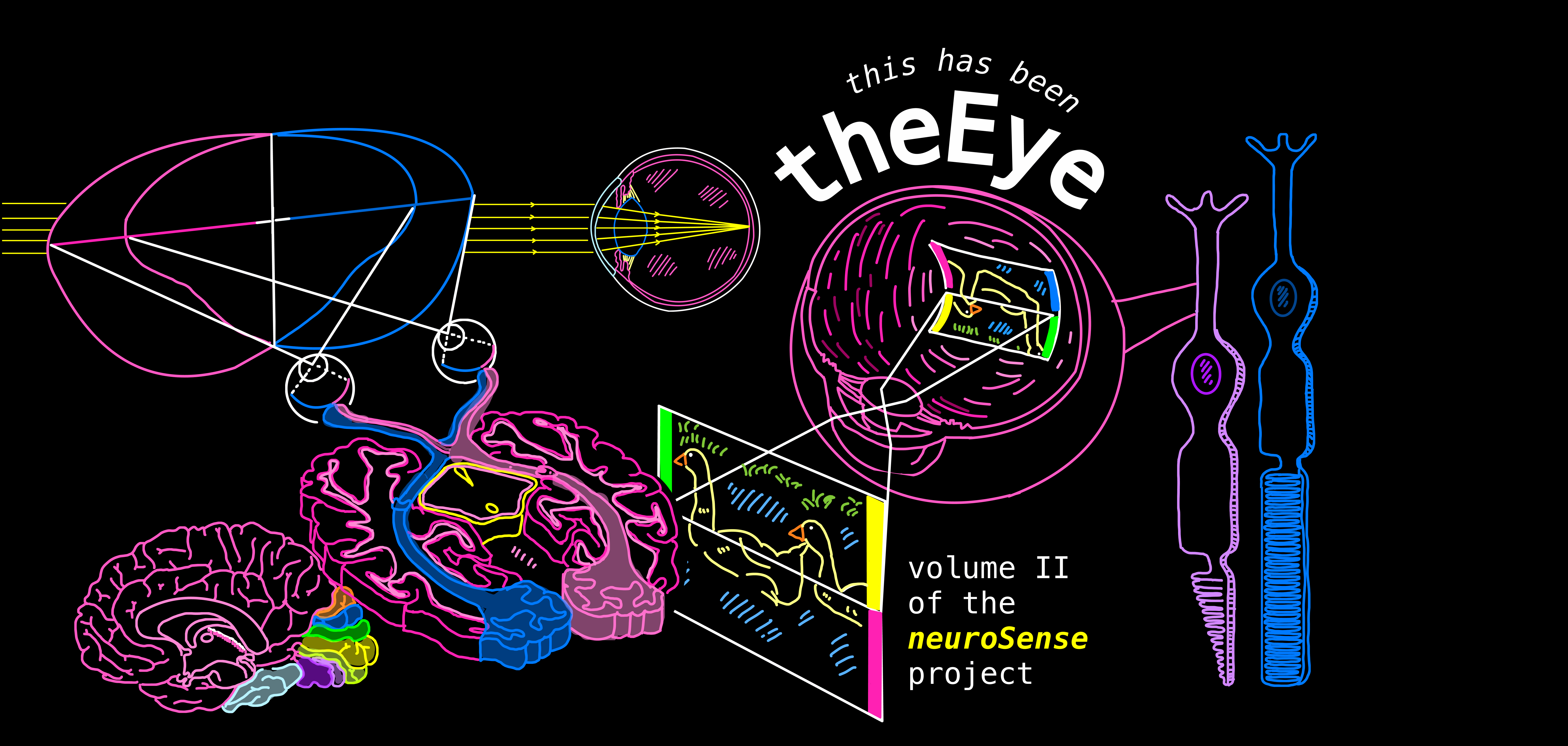
The Central Visual System

Ganglion cells signal the differences in brightness. By responding strongly to contrasts between light and dark, they highlight the edges, contours, and transitions in our visual field.
This contrast-based coding is what allows the brain to detect shapes, boundaries, and fine details in the world around us.
Colour-Opponent Processing
These are largely P-type cells, which are small, wavelength-sensitive, and function as a subtype of on-center cells. They compare activity from different cones to encode colour contrast.
Let’s take red–green opponent cells as an example (see top left & right). These cells compare inputs from red- and green-sensitive cones and respond in opposite ways depending on where the light lands:
1. Green light in the center of the receptive field (-M) decreases firing, while red light in the surround increases it (+L). Net effect depends on which region is stimulated, but the opponency is clear.

You can perceive blue + red together. You can perceive blue + green together. But you cannot perceive red + green in the exact same place at the same time — your visual system resolves this combination as yellow instead.
The same occurs for the blue–yellow opponent pair: you cannot perceive blue + yellow simultaneously in the same spot. When both pathways are activated together, the brain cancels them out, and the result is a perception closer to white or grey, depending on intensity.
Why this matters...
When light strikes the surround, it typically produces inhibition. When that inhibition stops (for example, when you look away), the system briefly rebounds in the opposite direction.
This rebound is what creates the American flag afterimage illusion (that you learnt in Part I!). Staring at yellow stimulates one pathway strongly, and when that stimulation ends, the opposing pathway briefly fires more — making you perceive blue!
The Retinofugal Projection
Once signals exit the eye, they follow a specific pathway called the retinofugal projection — “retino” (retina) + “fugal” (to flee). In other words, it’s the route that visual information takes as it leaves the retina and travels deeper into the brain.
There are three major structures in this pathway.
- Optic Nerve: The axons of the ganglion cells bundle together to form the optic nerve, carrying information out of each eye.
- Optic Chiasm: This is where some of those axons cross (decussation) to the opposite side of the brain.
- Optic Tract: After the chiasm, the reorganised bundle of axons continues deeper into the brain toward targets like the LGN and superior colliculus.

- Information from the left visual field goes to the right hemisphere.
- Information from the right visual field goes to the left hemisphere.

But the crossing is not complete — only the axons from the nasal half of each retina cross. The temporal half stays on the same side (ipsilateral).
This is why each hemisphere receives information from both eyes, but only from one visual field.
This arrangement tells us how the eyes capture visual information AND how the brain organises it so both hemispheres get a complete map of the opposite side of the world.

Note: We use nasal to describe when something is closer to your nose, and temporal when something is closer to the sides of your head (behind your ears).
Visual Field
Each eye captures both sides of the visual field, but different halves of the retina handle different parts.
- The nasal retina of each eye captures the outer part of the visual field (and its axons cross at the optic chiasm).
- The temporal retina captures the inner part (and its axons stay ipsilateral).
Binocular Visual Field
The binocular visual field is the region of the visual world that both eyes can see at the same time. This overlap exists because of our inverse topographic arrangement — the left half of each retina sees the right half of the world and vice versa.

Lesions along the retinofugal pathway produce very predictable visual deficits because each structure carries specific parts of the visual field. Let’s walk through the major ones.
- Transection of left optic nerve
All input coming from the left eye is lost.

- The right eye still captures both its peripheral field and the central (binocular) region, so visual information is still reaching the brain from those areas.
This means vision from the right visual field and the shared central field is preserved, even though the left eye is completely nonfunctional.
Vice versa occurs with the transection of the right optic nerve.
- Transection of left optic tract
All information from the right visual field is lost.

- The left optic tract ONLY carries signals representing the right side of the visual world — from BOTH eyes.
Once that tract is cut, nothing from the right visual field can reach the left hemisphere. But, vision from the left visual field is preserved because the right optic tract is still intact.
- Transection of the optic chiasm
All peripheral vision is lost, but the binocular visual field is preserved.

- Information from the peripheral visual field lands on the nasal retina of each eye. The nasal retinal axons are the only ones that cross at the optic chiasm. If the chiasm is cut, these crossing fibres can’t reach the opposite hemisphere anymore → thus, peripheral vision is lost on both sides.
- Meanwhile, the binocular visual field lands on the temporal retina, and temporal fibres do NOT cross. Because they stay on the SAME side, they are unaffected by the lesion → thus, the brain still receives central visual information.
Thalamic and Non-Thalamic Targets of the Optic Tract

This layered structure reflects a segregation of visual input by eye:
- Layers 2, 3, and 5 receive information from the ipsilateral (same) eye.
- Layers 1, 4, and 6 receive information from the contralateral (opposite) eye.

So, different pieces of information about where light is coming from are routed to different layers of the LGN.
We don’t fully know why the LGN is structured this way but there’s a leading hypothesis: this organisation preserves the origin of visual information (which eye it came from, and which part of the retina), while still allowing that information to be compared and integrated later on.
Thus, the LGN both separates input by eye and preserves information about which retinal ganglion cells are sending the signal as it travels through the optic nerve, optic chiasm, and optic tract.
LGN Layer Organisation
- M-Type (Magnocellular) cells: These mainly respond to light-dark contrast (ON/OFF center organisation). They are big cells and have large receptive fields - good for detecting movement, broad shapes, and fast changes in luminance.
- P-Type (Parvocellular) cells: These often show colour-opponent responses. They are small cells, have small receptive fields, and are sensitive to different wavelengths - important for colour and fine detail.

Layers 1-2 → receive input from M-type ganglion cells. These are the magnocellular layers (magno = big).
Layers 3–6 → receive input from P-type ganglion cells. These are the parvocellular layers (parvo = small).
K1–K6 → koniocellular layers tucked between them.
By keeping these streams separate, the LGN sends an incredibly detailed, structured report forward into the visual cortex. This organisation lets the brain analyse motion, colour, and form in parallel without losing any information from either eye or either cell type.
Non-Thalamic Targets of Retinal Ganglion Cells
All of the following structures receive direct input from the retina.
- Hypothalamus (Suprachiasmatic Nucleus – SCN): Regulates our circadian rhythms - using light-dark information to coordinate daily cycles.
e.g., sleep, metabolism, hormone release, and body temperature.
- Pretectum: Controls pupillary light reflex and lens accommodation. When light increases, the pretectum activates parasympathetic (rest and digest) pathways, which constricts our pupils. This helps maintain appropiate focus and retinal illumination.
- Superior Colliculus: Coordinates orienting movements of the eyes, head, and neck - integrating visual input with other sensory modalities to guide attention toward stimuli.
Note: Saccades are rapid, ballistic eye movements that shift the focus of gaze from one point to another.
This is essential for scanning scenes and tracking fast-moving objects. Even when reading or looking at a still scene, your eyes make small involuntary saccades to build a complete image.
The Primary Visual Cortex

V1 sits along the calcarine fissure in the occipital lobe and begins transforming basic retinal input into more complex representations such as edges, orientations, and motion direction.
Key Features of V1
- Retinotopy: Neighbouring areas of the retina feed neighbouring areas in the LGN and V1 — the visual system preserves spatial organisation of the image.
- Ganglion cells carry information about colour (from P-type cells) and light/dark contrast
(from M-type cells).
This information is relayed into V1, where neurons are also organised in layers that process different aspects of the visual signal.
Foveal (central) vision is overrepresented in V1. Many more retinal ganglion cells represent the center of the visual field compared to the periphery. As a result, a large part of V1 is devoted to high-acuity central vision and only a small portion to coarse peripheral vision.

- Lamination: The LGN is organised into layers — and V1 is layered too!
- When we look at V1, we see that different layers are specialised for different stages of input and output processing. This layered structure helps preserve and organise visual information as it moves through the cortex.
Information coming from the LGN
Input from LGN layers 1–2 projects mainly to layer IV-Cα in V1.
Input from LGN layers 3, 4, and 6 projects mainly to layer IV-Cβ.
Layer IV-C is the primary input layer of V1 — this is where neurons first receive axons coming directly from the LGN.

- Ocular Dominance: Ocular dominance refers to the fact that we often rely more on one eye than the other — for example, having better acuity in one eye or finding it easier to fixate on objects with a particular eye.
- In V1, information from each eye initially arrives segregated. That is, signals from the left and right eyes are kept separate when they first enter the cortex.
As this information moves into more superficial layers of V1, the signals from both eyes begin to mix.
- V1 initially preserves information about which eye a signal came from.
- But at higher levels of processing, that information is integrated — which is exactly what we want for unified visual perception.
-
As a result, some neurons respond more strongly to input from the left eye, some to the right eye, and others to a combination of both.

New Types of Receptive Fields
Because P-type cells project mainly to layers 3, 4, 5, and 6, and M-type cells project mainly to layers 1 and 2, this layered organisation in V1 preserves many of the same receptive field properties we see earlier in the LGN.

Orientation & Direction Selectivity
Orientation Selectivity
Neurons are still often dominated by input from one eye, but they now respond to information from both eyes, reflecting the beginning of binocular integration.
Some neurons no longer respond best to circular spots of light. Instead, they fire most strongly when a stimulus with a specific orientation (for example, a vertical or horizontal bar of light) appears in their receptive field. These neurons are therefore orientation-selective.


Other neurons go a step further and are also sensitive to motion direction.
They fire more when a light stimulus moves through their receptive field in a particular direction, but respond weakly or not at all if the same stimulus moves in the opposite direction.
Theoretical Cortical Modules
Neurons within the same column tend to respond best to the same orientation of a stimulus (for example, vertical vs. horizontal edges).
These column-based groupings are often referred to as theoretical cortical modules — functional units that process specific visual features like orientation, eye of origin, and spatial location.

Damage to different regions of V1 leads to characteristic visual deficits, depending on the size and location of the lesion.
- Hemianopia: The complete loss of vision in one half of the visual field.
For example, a lesion in the left visual cortex results in loss of vision in the right visual field (the contralateral visual field).
This affects the right half of the visual field from both eyes, not just one eye.
- Scotoma: A scotoma results from a small, localised lesion affecting a group of neurons in V1.
This produces a blind spot in a specific region of the visual field rather than loss of an entire half. The size and location of the scotoma directly map onto the damaged cortical region due to V1’s retinotopic organisation.

What happens next is less understood, but we have a strong working model.
Once visual information reaches V1, processing is far from over. Beyond V1 lies the extrastriate cortex, a collection of 20+ specialised visual areas, including: V2, V3, V3A, V4, V5 (also called MT), and higher-level object and face recognition regions.

Retina → LGN → V1 → Extrastriate cortex
Processing Streams
- Dorsal Stream (“Where / How” Pathway): Specialises in motion, spatial location, and visually guided action.
i.e., Where is it? How is it moving? How do I interact with it?
- Area MT / V5: Dedicated to motion perception. Neurons here respond strongly to direction and speed of movement.
- Akinetopsia: Results from damage to V5. Vision is otherwise intact, but motion perception is lost. The world appears as a series of static snapshots rather than smooth movement.
e.g., A moving car is perceived as suddenly appearing closer, without visible motion between positions.

- Ventral Stream ("What" Pathway): Specialises in object identity, shape, and colour. i.e., What is it?
- Area V4: Processes colour and object form.
- Achromatopsia: Damage to V4. Loss of colour perception (world appears in shades of grey)
- Object Agnosia: Patients can see, reach, and copy objects. But they cannot recognise or identify what the objects are.
- Face-Specific Processing: A specialised extension of the ventral stream dedicated to facial recognition.
- Fusiform Face Area (FFA): Contains neurons that respond more strongly to faces than to other objects (e.g., houses).
- Prosopagnosia (face blindness): Can be acquired due to brain damage or developmental. Individuals with prosopagnosia cannot recognise faces, even though vision is intact and object recognition may still be normal. They often rely on alternative cues (voice, posture, hairstyle) to identify people.
Short answer: yes.
- Prosopometamorphopsia (PMO): This is an extremely rare disorder involving distorted perception of faces. Distortions are often face-specific, while other objects appear normal.
This is usually caused by right-hemisphere lesions in the ventral occipito-temporal cortex, near face-selective regions like the fusiform gyrus, disrupting how facial features are integrated rather than how they are seen.
- PMO Case Example: A patient reported seeing demon-like facial distortions in everyone he encountered. The distortions appeared regardless of where the face was in the visual field. Strikingly, the distortions occurred in real life but not when viewing faces on screens.
From the retina and LGN, where information is filtered and organised, through V1’s feature extraction and into the extrastriate cortex, visual perception emerges through progressively abstract representations.
Damage at different stages does not simply reduce vision, but alters what is perceived — motion, colour, objects, and faces.
Disorders such as prosopometamorphopsia highlight this principle especially clearly: when non-primary visual regions are disrupted, basic sight may remain intact while the brain’s interpretation of visual information becomes distorted.
This shows us that what we “see” is the brain’s best reconstruction of reality, shaped by neural specialisation, integration, and prior organisation.
Summary
- Ganglion cells emphasise contrast, not just absolute brightness.
They fire differently depending on light in the center vs. surround, helping the brain detect edges, contours, and boundaries.
- Colour-opponent processing organises colour perception.
Some ganglion cells compare wavelengths (e.g., red vs. green, blue vs. yellow).
Activating one suppresses the other — which explains colour after-images and why red-green isn’t perceived simultaneously in one spot (resolves to yellow).
- Retinofugal projection carries signals from eye → brain.
Visual information leaves via the optic nerve, partially crosses at the optic chiasm, and continues through the optic tract.
- Partial decussation (“crossing”) organises visual fields.
Left visual field → right hemisphere.
Right visual field → left hemisphere.
The overlapping binocular field is seen by both eyes.
- Lesions along the pathway predict specific vision loss.
Optic nerve cut → vision lost in that eye.
Optic tract cut → vision lost in the opposite visual field.
Optic chiasm cut → vision lost in the peripheral (“tunnel vision”) but central vision preserved.
- The LGN (a thalamic target) organises visual information into layers.
Each layer receives input from a specific eye and a specific type of retinal ganglion cell, keeping signals neatly separated before they reach the cortex.
M-type (magnocellular) cells: Specialise in motion and contrast.
P-type (parvocellular) cells: Specialise in colour and fine detail.
- Non-thalamic targets support reflexive and biological functions.
Hypothalamus (circadian timing), pretectum (pupil reflex), and superior colliculus (eye-movement orienting).
- The primary visual cortex (V1) maps the world precisely.
Neighbouring retinal areas map to neighbouring cortex (retinotopy), with extra representation for the fovea.
- V1 is layered and mixes inputs gradually. Early layers keep left/right eye separate; higher layers combine them, forming ocular dominance columns.
- New receptive fields emerge in V1. Neurons become selective for orientation (edges at angles) and direction of motion, laying groundwork for shape and movement perception.
- Damage to V1 causes predictable deficits.
Large lesions → hemianopia (half-field blindness).
Small lesions → scotomas (blind patches).
- Beyond V1, vision splits into two major streams.
Dorsal stream (“where/how”): Motion, movement, spatial mapping.
Ventral stream (“what”): Object identity, colour, faces.
- Specialised cortical regions handle specific visual roles.
V5/MT: Motion (damage → akinetopsia, motion blindness).
V4: Colour/form (damage → achromatopsia).
Fusiform Face Area: Faces (damage → prosopagnosia, face blindness).
- Some disorders come from lesions outside primary vision areas.
Prosopometamorphopsia (PMO) causes distorted faces, likely from ventral visual stream damage, even though basic sight remains intact.
Key Terms
Below are definitions of common terms used throughout the volume.
Neurons
Neurons are the basic cells of the nervous system.
They send and receive information through electrical and chemical signals.
- Cell body (soma): The “hub” of the neuron. It contains the nucleus and all the usual cell machinery needed to stay alive.
- Neurite: A collective term to denote both axons and dendrites.
- Axons allow a neuron to pass information to other neurons.
Dendrites allow a neuron to receive information from other neurons.


Action Potential
A rapid electrical “spike” that neurons use to send information. It’s an all-or-nothing event: once the signal starts, it travels down the axon until it reaches the end of the cell, where it triggers the release of neurotransmitters.
An all or nothing approach is like flushing a toilet! You can either flush (have an action potential), or not flush (not have an action potential).
It doesn’t matter how hard/long you push the handle, same functionality applies.
These are the brain’s chemical messengers. They carry signals between neurons across the synapse (the tiny gap between cells).
When an action potential reaches the end of an axon, it triggers the release of neurotransmitters. These molecules cross the synapse and bind to receptors on the next neuron, influencing whether that cell will fire its own signal.
Different neurotransmitters have different effects: some excite (make a neuron more likely to fire), others inhibit (make it less likely).
GABA (gamma-aminobutyric acid) is the main inhibitory neurotransmitter. It calms neural activity and prevents circuits from becoming over-excited (too much unchecked exicatory activity can actually damage our neurons).
Together, glutamate and GABA create a precise push-and-pull balance that allows the visual system, and the entire brain, to remain stable, responsive, and incredibly efficient.
Neurons rely on gradients to generate signals. A gradient simply means there is more of something on one side of the cell membrane than the other.
Chemical gradient: There are different concentrations of ions (like sodium and potassium) inside vs. outside the neuron. Ions naturally want to move from areas of high concentration to low concentration due to entropy. This is called diffusion.

The neuron's interior is typically negatively charged relative to the outside. Positively charged ions are attracted to negative areas and repelled by positive ones, creating an electrical force that can either reinforce or oppose the chemical gradient.

When the chemical and electrical gradients work in the same direction, ions flow easily through open channels.
When they oppose each other, the ion may reach equilibrium (where the two forces balance out) or require energy to move against the combined gradient. This stored electrochemical energy is what neurons use to generate and propagate signals.
In photoreceptors, these gradients determine whether ion channels open or close, which controls whether the cell is depolarised or hyperpolarised when light hits the retina.
Remember, neurons maintain a difference in charge between the inside and outside of the cell. The inside is usually more negative than the outside (this is called the resting membrane potential).
- Depolarisation: The inside becomes less negative (more positive) moving closer to the threshold needed to fire an action potential. This increases the likelihood that an action potential will occur.
- Hyperpolarisation: The inside becomes even more negative, moving further from the threshold. This reduces the chance that an action potential will occur.
Neurons respond to neurotransmitters using different kinds of receptors.
Ionotropic receptors: These are fast, direct receptors. When a neurotransmitter binds, the receptor opens an ion channel immediately, allowing ions to flow across the membrane and quickly change the cell’s electrical state. Think of ionotropic receptors as light switches — almost instant on/off.
Metabotropic receptors: These are slower, indirect receptors. Instead of opening a channel right away, they trigger a biochemical cascade inside the cell that eventually alters ion channels or cell behaviour. Think of metabotropic receptors as dimmer switches — slower, but capable of fine control.
Thus, different neurons can respond very differently to the same neurotransmitter.
Together, these two systems let the visual system be both fast and highly adaptable.
Directional Terms
When describing the brain, we use special anatomical directions to keep things consistent.
e.g., the frontal lobe is anterior to the visual cortex.
Posterior: Toward the back.
e.g., the cerebellum is posterior to the visual cortex.
Dorsal: Toward the top.
e.g., In the brain, toward the top of the head.
Ventral: Toward the bottom.
e.g., In the brain, toward the base of the skull.


Medial: Toward the midline.
e.g., middle of the body/brain.
Lateral: Toward the sides.
e.g., your ears are lateral to your eyes.
Rostral: Toward the nose
(or front of the brain).
Caudal: Toward the tail
(or back of the brain).
Ipsilateral: Same side.
Contralateral: Opposite side.
e.g., The visual cortex is superior to the brainstem.
Inferior: Below.
e.g., the brainstem is inferior to the visual cortex.
Closing Remarks
Light must be captured, refracted, inverted, filtered, encoded, and constantly corrected by networks of cells that evolved to extract just the information we need to survive.
Vision is less a mirror of reality and more a sophisticated model of it — assembled in real time from limited data. And yet, this imperfect system lets us read, recognise faces, navigate the world, appreciate art, and experience sunsets!
Vision is powerful not because it is complete, but because the brain turns the messy, chaotic input of reality into something we can understand - providing yet another example of how spectacular the brain truly is.
Stay tuned! The next neuroSense volume will turn from the eye to the nose and mouth, exploring how we perceive the world through our chemical senses.
thanks for reading!
amethyst